
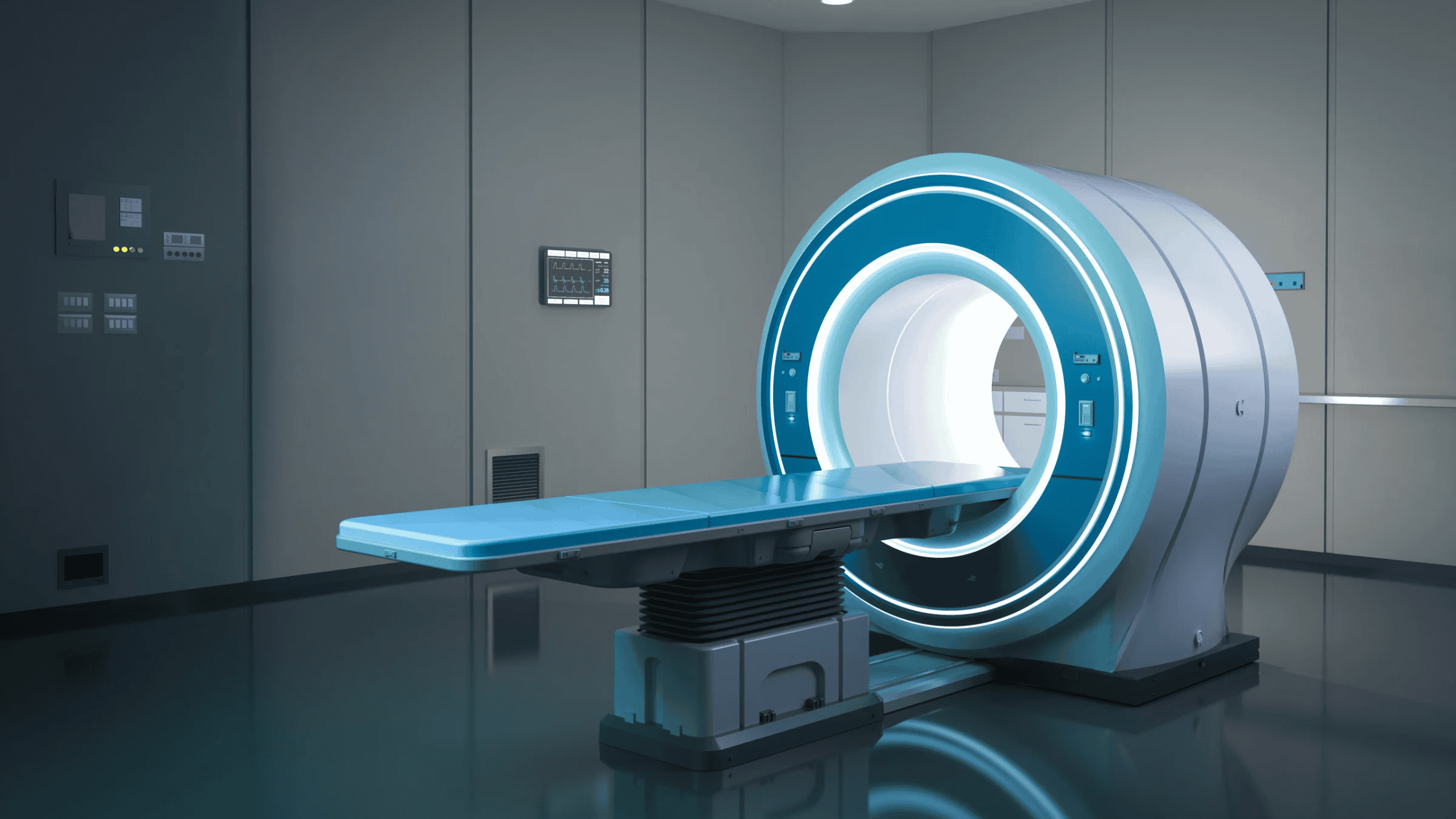
When back or neck pain persists, determining the underlying cause can require more than a physical examination. Magnetic resonance imaging, commonly called an MRI, can provide detailed images of the spine and the soft tissues surrounding it. Unlike an X-ray, which primarily shows bones, an MRI can help physicians evaluate discs, nerves, the spinal cord, ligaments and other structures.
Understanding what a spinal MRI can reveal may help patients better understand why imaging is recommended and how the results can influence treatment decisions.
What Is a Spinal MRI and How Does It Work?
A spinal MRI is an imaging test that uses a strong magnetic field and radio waves to create detailed pictures of the spine. A spinal MRI does not use ionizing radiation, unlike an X-ray or CT scan.
During a spinal MRI, the patient lies on a movable table that slides into the MRI scanner. The machine creates images in multiple planes, allowing physicians to examine the spine from different angles. Depending on the reason for the examination, an MRI may focus on the cervical spine, thoracic spine or lumbar spine.
What Can an MRI Reveal About Spinal Discs?
An MRI can provide a detailed view of the discs located between the vertebrae. These discs act as cushions that help absorb forces and allow the spine to move.
An MRI can reveal changes such as disc degeneration, disc bulges and herniated discs. Disc degeneration can involve changes in the disc’s structure and hydration, while a herniated disc occurs when disc material extends beyond its normal boundaries.
These findings can sometimes help explain back or neck pain, particularly when a disc problem is affecting nearby nerves. However, disc abnormalities can also occur in people who have no symptoms, so MRI findings must be considered alongside the patient’s symptoms and physical examination.
Can an MRI Show a Pinched Nerve?
An MRI can show whether structures in the spine may be compressing or irritating a nerve. Nerve compression can occur when a herniated disc, bone spur, arthritis-related changes or narrowing of the spinal canal reduces the space available for a nerve.
When a nerve is compressed, symptoms may extend beyond the back or neck. Depending on the location of the affected nerve, patients may experience radiating pain, numbness, tingling or weakness in an arm or leg.
For patients with symptoms such as sciatica or cervical radiculopathy, an MRI can help identify structural changes that may correspond with the affected nerve.
What Does an MRI Show About Spinal Stenosis?
An MRI can provide valuable information about spinal stenosis, a condition involving narrowing of the spaces within or around the spine. An MRI can show narrowing of the central spinal canal as well as the openings where spinal nerves exit the spine.
Spinal stenosis can develop as the spine changes with age. Disc degeneration, arthritis, thickened ligaments and bone overgrowth can all contribute to narrowing.
An MRI can help physicians determine where narrowing occurs and whether nerves or the spinal cord appear to be affected. These findings can be particularly useful when evaluating symptoms such as leg pain, numbness, weakness or difficulty walking.
Can an MRI Detect Problems With the Spinal Cord?
An MRI is particularly useful for evaluating the spinal cord and surrounding soft tissues. An MRI can reveal abnormalities involving the spinal cord that may not be visible on standard X-rays.
Depending on the circumstances, MRI findings may help identify inflammation, compression, certain tumors, infections, traumatic injuries or other abnormalities affecting the spinal cord. MRI may also be used to evaluate conditions involving the spinal cord when symptoms include unexplained weakness, sensory changes or coordination problems.
Can an MRI Show Arthritis and Other Changes From Aging?
An MRI can reveal several structural changes associated with spinal arthritis and aging. These changes may include cartilage and joint deterioration, bone spurs, disc degeneration and thickening of supporting ligaments.
These findings are common as people get older, and an MRI abnormality does not automatically mean that it is the source of a patient’s pain. The significance of an MRI finding depends on its location, severity and relationship to the patient’s symptoms.
This is why spinal imaging is most useful when interpreted in the context of a complete clinical evaluation.
Does Everyone With Back Pain Need an MRI?
Not everyone with back pain needs an MRI. Many episodes of back pain improve with conservative treatment and do not require advanced imaging.
An MRI may be considered when symptoms persist despite appropriate treatment, when significant nerve symptoms are present, or when a physician needs more information to evaluate a suspected spinal condition. MRI may also be appropriate when certain warning signs raise concern for a more serious underlying problem.
The decision to order an MRI depends on the patient’s symptoms, medical history, examination findings and treatment needs.
How Can MRI Results Affect Spine Treatment?
MRI results can help physicians connect a patient’s symptoms with specific structural findings. An MRI may help determine whether treatment should focus on physical therapy, targeted injections, activity modification or another nonsurgical approach.
In some cases, MRI findings may also help determine whether a patient should be evaluated for a surgical procedure. However, treatment decisions should not be based on an MRI image alone. The goal is to understand how the imaging findings relate to the patient’s pain, function and overall clinical picture.
What Should Patients Remember About Spinal MRI Results?
A spinal MRI provides a detailed picture of the structures inside and around the spine, but the picture is only one part of the diagnostic process. An MRI can reveal disc problems, nerve compression, spinal stenosis, spinal cord abnormalities, arthritis-related changes and other structural findings.
Most importantly, an abnormal MRI does not necessarily mean that a patient needs invasive treatment. The most meaningful interpretation comes from combining imaging findings with symptoms, physical examination results and functional limitations. This comprehensive approach can help identify the likely source of spinal pain and guide an appropriate treatment plan.